
Minimal Radiation Exposure, Maximum Image Details
Since Wilhelm Conrad Röntgen immortalized his wife’s hand in the first image obtained using X-rays, a great deal of progress has been made. The technology now not only takes into account two, but three dimensions: Today, 3D X-ray imaging has established itself as an indispensable tool for answering complex clinical questions. These three-dimensional images of the body regions to be examined are created by exposing them to radiation using an X-ray system. To date, the entire region of the body has to be uniformly exposed to radiation – even though in most cases, only a small part of the image volume obtained is relevant for answering the clinical question. Is it technically and economically possible to design the imaging components’ guidance systems in such a manner that only the required regions are exposed to radiation?
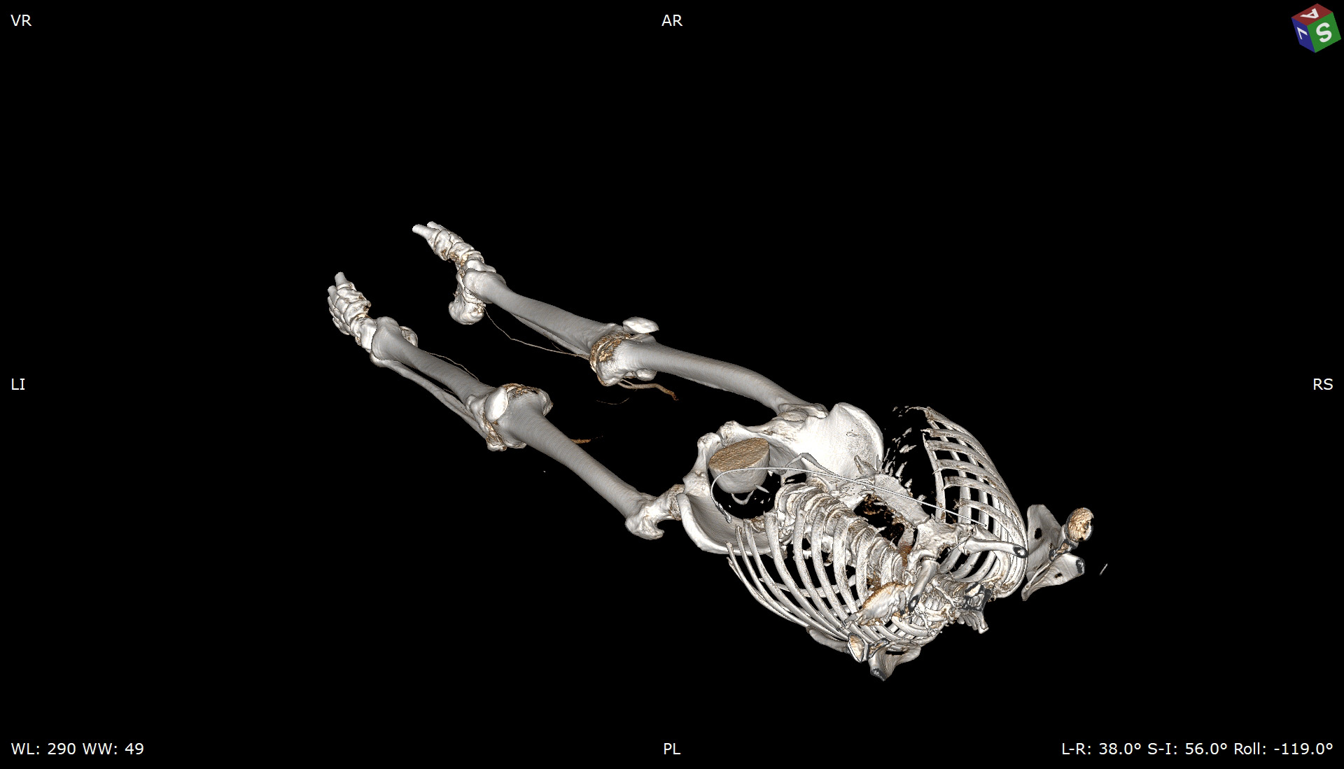
3D X-ray image
Each type of tissue is different
Different tissue types in the body react to X-radiation with varying degrees of sensitivity. Particularly critical, for example, are the lenses of the eye, glandular tissue such as the breast and thyroid glands, reproductive organs, and the intestines. Radiologists generally avoid X-raying these anatomical structures, as radiation-induced genetic alterations in these structures may have particularly severe repercussions. Accordingly, an equal dose with weighted distribution throughout the tissue dose is aimed for. The total dose of radiation administered remains the same, but the more sensitive tissue types are significantly less irradiated. 3D X-ray imaging systems available on the market today are unable to meet these complex medical requirements.
The imaging components of a 3D X-ray system – the X-ray source and X-ray detector – are guided mainly along standardized, circular trajectories. If it were possible to better adapt the motion paths of these components to each individual case, the reduction of radiation exposure of sensitive structures such as reproductive organs could be achieved, while at the same time maintaining most of the image quality. As part of his dissertation at Fraunhofer IPK, medical engineer Felix Fehlhaber is therefore investigating how such specialized motion paths can be implemented in practice.
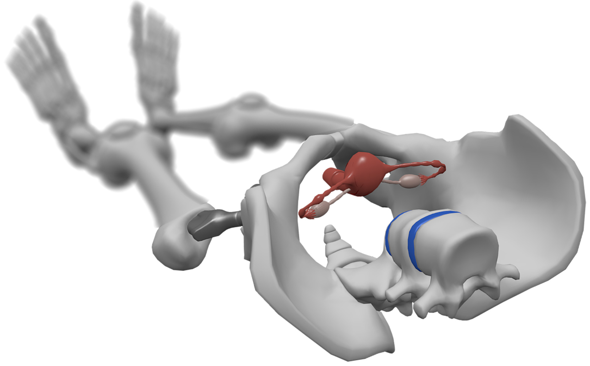
Model of a pelvis with X-ray sensitive reproductive organs
The proof is in the simulation
For this purpose, he has developed simulation software that simulates medical X-ray processes in a physically precise manner. »The method is based on the Monte Carlo principle: A large number of small experiments are used to approximate the actual result,« explains Fehlhaber. The experiment simulated in this case: A photon, which is a light particle operating in the X-ray spectrum, is followed virtually on its path through realistically modeled tissue. At the same time, all medically relevant physical interaction effects are taken into account. Fehlhaber explains: »This provides a highly precise overview of where photons release the most energy in the body – i.e., potentially causing damage due to radiation – and how photons propagate in the body.« Utilizing special quality assessment procedures, he is also able to objectively evaluate how much a modified arrangement of imaging components in the simulation would affect the quality of the actual image.
Based on findings from the simulation experiments, Fehlhaber expanded the software to include an optimization algorithm. This algorithm identifies the optimal trajectories for specific clinical issues. It automatically determines which path the imaging components will need to follow to achieve an optimal balance between the lowest possible radiation exposure and adequate image quality for the anatomical structures being examined. Fehlhaber was able to prove that this method can reduce exposure to radiation. Above all, highly sensitive structures such as the uterus and ovaries are exposed to significantly less radiation. »However, the exact simulation of all fluoroscopies required to optimize the trajectory necessitates a great deal of computing power. For complex tissues, this currently takes several days,« explains Fehlhaber. He will be publishing the results of his experiments as part of his doctorate in the fall of 2021.
Back to the future
Considering the current state of the art and the hardware developments over the past decade, it can be assumed that such optimizations will only become established in the field of radiology in a few years. Only then will the original goal of reducing the effective radiation dose become ready for mass implementation. Until such time, researchers at Fraunhofer IPK will continue to develop and optimize the speed of the algorithm. Fehlhaber envisions a great deal of potential for future research questions: »The medical models which are indispensable for realistic simulations are constantly being refined. And we are also using the simulation software to investigate procedures that improve the image quality of 3D X-ray imaging.«
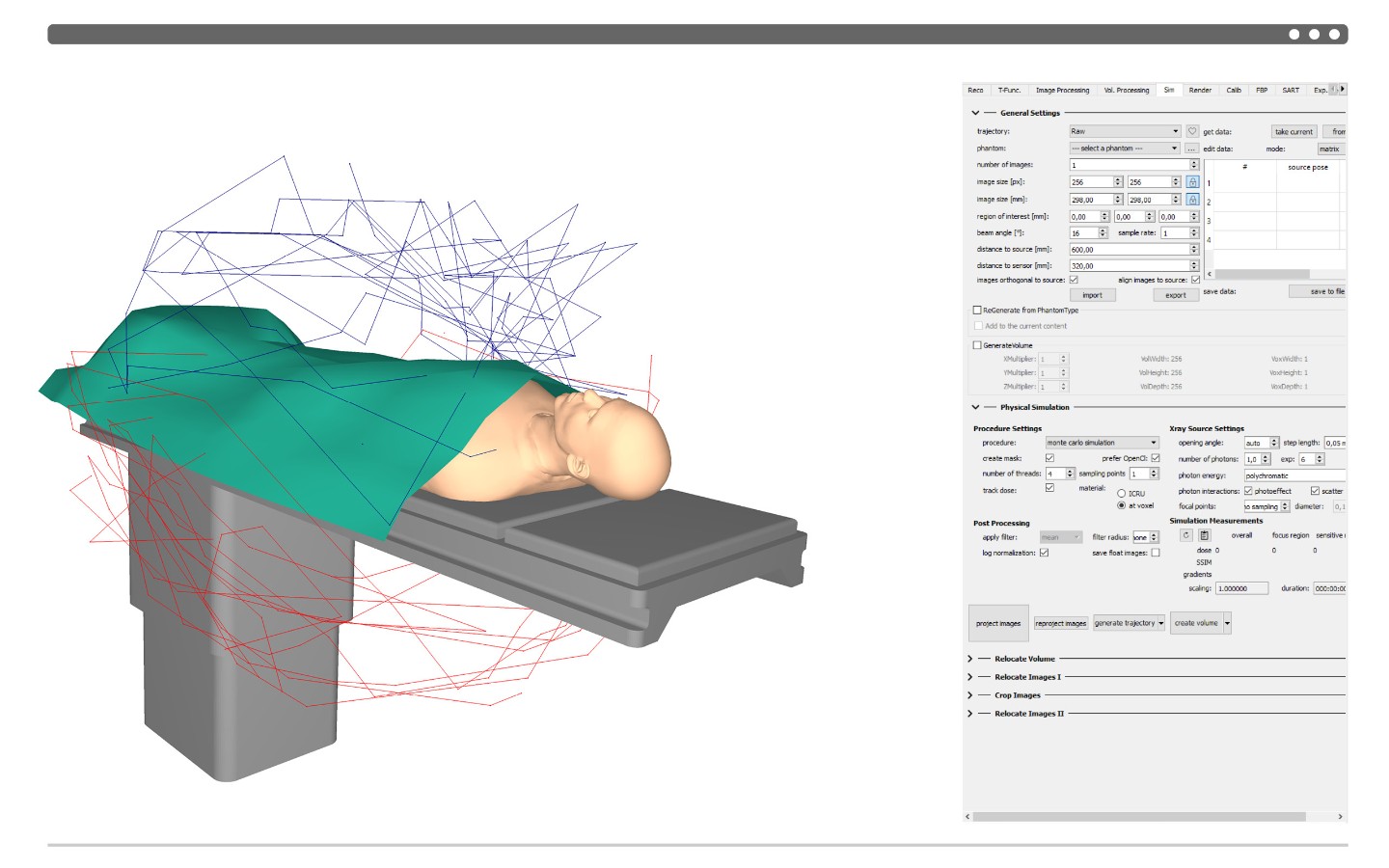
Simulated trajectory of X-ray source (red) and detector (blue) with significantly reduced effective dose during scan of the pelvis